At a Glance
Chiari malformation is a structural condition in which parts of the brain (typically, the cerebellum) extend downward into the spinal canal, sometimes compressing the brainstem that controls breathing and sleep. This compression can increase the risk of sleep apnea, particularly central sleep apnea, and treatment may involve monitoring symptoms, sleep studies, CPAP or similar therapies, or surgery to relieve pressure on the brain.
The brain is a complex organ that controls movement, processes sensory information, and regulates essential functions like breathing and sleep. Chiari malformations are typically caused by structural changes in the skull and brain that occur before birth.
In this condition, parts of the brain — specifically, the brain stem and cerebellum — are displaced from their normal position. When these structures shift, they can place pressure on the surrounding areas and disrupt normal breathing patterns, increasing the risk of sleep apnea.
In this article, we'll explain how Chiari malformations affect the brain and airway, the symptoms to watch for, and how they're diagnosed and treated.
Could Your Symptoms Be Sleep Apnea? Find Out
Some breathing issues are connected to sleep apnea. Sleep Doctor’s home sleep study helps you get clear answers without a lab visit.

What Is Chiari Malformation?
Chiari malformations occur when the back of the skull forms an abnormal shape, forcing the lower portion of the brain called the cerebellum towards the base of the skull. This can push the cerebellum into the foramen magnum, the opening where the brain stem meets the spinal cord.
There are several subtypes of Chiari malformations that vary in degree of severity. Type 1 and 2 Chiari malformations are the most common. Each type is classified by which parts of the brain are pushed below the base of the skull and what kind of symptoms an individual experiences.
Type 1 Chiari Malformation
In a type 1 Chiari malformation (CM), the lowest lying part of the cerebellum descends through the opening in the base of the skull and into the spinal cavity. The brain stem remains inside of the skull.
In many people with this condition, the cerebellum may extend up to 3 millimeters into the spinal cavity and still be considered normal. A diagnosis of Chiari malformation is usually made when the cerebellum extends at least 5 millimeters or more past the foramen magnum.
Type 2 Chiari Malformation
In type 2 Chiari malformation, also called Arnold-Chiari malformation, more parts of the brain are displaced and pushed down through the base of the skull. The middle part of the cerebellum, the lowest lying part of the cerebellum, and part of the brain stem protrude into the spinal cavity.
Type 2 CM is usually associated with a condition called myelomeningocele. In myelomeningocele, the bones of the spine don't fuse together completely and leave a gap. Through that gap, the spinal cord, cerebrospinal fluid, and spinal cord tissues (meninges) can bulge out, making them susceptible to damage.
Other Chiari Malformations
Less common types of Chiari malformations have also been identified. Types 3 and 4 involve more severe abnormalities of the brain and upper spine and are typically diagnosed early in life.
In some cases, healthcare providers may use the term Chiari malformation type 1.5 to describe features that fall between types 1 and 2 but occur without spinal defects. While this classification is not widely used, type 1.5 has been linked to a higher likelihood of obstructive sleep apnea in some individuals.
How Chiari Malformation and Sleep Apnea Are Connected
Adults and children with CM are more likely to be diagnosed with sleep apnea. Experts do not yet have a definite explanation for the link between Chiari malformations and sleep apnea. However, there are a few proposed reasons.
Sleep apnea is a sleep disorder that causes disrupted breathing during sleep. The two types of sleep apnea are obstructive sleep apnea and central sleep apnea.
- Central sleep apnea (CSA) occurs when the brain fails to signal the respiratory muscles in the body to breathe. In Chiari malformations, the brain structures are displaced downward, causing the brain stem and cranial nerves to be compressed. Researchers have suggested that compression in this area may also affect part of the brain that controls breathing, which in turn can increase the risk of central sleep apnea.
- Obstructive sleep apnea (OSA) is caused by a structural blockage of airflow in the mouth or throat. The tongue or soft palate of the mouth, for example, may become lax during sleep and collapse into the airway, causing interrupted breathing. People with CM may develop obstructive sleep apnea if the muscles of their mouth or throat are not functioning properly and block airflow during sleep.
Chiari Malformation Causes and Symptoms
Chiari malformations are typically linked to structural differences in the brain and skull, most often present at birth, though symptoms may not appear until later in life. These changes can affect how the brain sits within the skull and how fluid flows around the brain and spinal cord. As a result, people with Chiari malformation may experience a range of symptoms that vary in type and severity depending on the extent of the displacement and the areas of the brain involved.
Causes of Chiari Malformation
Chiari malformations usually are caused by abnormal development of the brain or skull while a baby is still in the womb. If CM forms before birth, it's known as a primary or congenital Chiari malformation. While the exact cause of congenital CM is not known, researchers have several theories, including:
- Genetic mutations
- Deficiency of folic acid, a type of B vitamin, during pregnancy
- Infection during pregnancy
- Exposure of fetus to hazardous chemicals or drugs
Less commonly, skull changes may occur after a person has been born, leading to what’s called a secondary or acquired Chiari malformation. The cause of acquired CM can usually be identified and may include:
- Traumatic head injury after birth
- Infection after birth
- Pressure in the brain due to a tumor or the build-up of cerebrospinal fluid
Symptoms of Chiari Malformation
Although type 1 CM often develops before birth, people with this condition usually don't start experiencing symptoms until they are adolescents or adults. Some may not have symptoms at all. On the other hand, type 2 CM is usually diagnosed before birth or soon after.
Symptoms of type 1 and type 2 Chiari malformations can vary from person to person, depending on what parts of the brain and spinal cord are affected:
- Headache and neck pain, especially after coughing, sneezing, or laughing
- Muscle weakness or paralysis
- Difficulty swallowing
- Dizziness and fainting
- Problems with hearing or balance
- Vomiting or drooling
- Breathing issues


How Chiari Malformation Is Diagnosed
Chiari malformations (CMs) are diagnosed by using neuroimaging technologies that create pictures of the anatomical structures in the brain.
- Magnetic resonance imaging (MRI): Chiari malformations are usually diagnosed using magnetic resonance imaging, which can detect whether the cerebellum or brain stem are in an abnormal position.
- Computed tomography (CT) scan: When MRI cannot be used, a CT scan is an alternative to view the anatomical structures in the brain.
- Cerebrospinal fluid flow imaging: This method can show any abnormalities in flow of fluid through the brain.
Additionally, the doctor may ask questions about what kind of symptoms a person with suspected CM is experiencing. If they are having trouble sleeping, the doctor may refer them to sleep study to monitor breathing and electrical activity of the brain during sleep.
Treatments for Sleep Apnea With Chiari Malformations
There are several methods to treat sleep apnea associated with Chiari malformations. The specific treatments used depend on whether a person has central sleep apnea or obstructive sleep apnea.
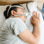
- Continuous positive airway pressure (CPAP): CPAP uses a machine to pump air into the nose and/or mouth during sleep in order to keep the airway open. CPAP provides an unchanging, set level of air pressure throughout the night. It is used to treat both central and obstructive sleep apnea.
- Bilevel positive airway pressure (BiPAP): Like CPAP, BiPAP pumps air into the airway during sleep, but BiPAP differs from CPAP in that the level of air pressure for breathing in is different from the level of air pressure for breathing out. It is primarily used in central sleep apnea treatment.
- Adaptive servo-ventilation (ASV): ASV is a breathing device worn by some people with central sleep apnea. The device provides air pressure to assist in breathing during sleep and continuously adjusts the air pressure based on a person’s breathing patterns.
- Supplemental oxygen: Some people with central sleep apnea benefit from breathing extra oxygen at night, either on its own or in combination with CPAP therapy.
Decompression surgery may also be an option for individuals who have Chiari malformations that are causing symptoms. Surgical procedures may improve the flow of cerebrospinal fluid and lessen the pressure on the head.
However, decompression surgery as a treatment for sleep apnea in people with CM has not been thoroughly studied. In some case reports, surgery improved some symptoms of sleep apnea, but additional treatment was needed after surgery.
Frequently Asked Questions
Chiari malformation is considered relatively uncommon, but exact rates can vary depending on how it's defined and diagnosed. Recent data suggest that it’s present in between 1% and 4% of people. With the increased use of imaging tests like MRI, more cases, especially mild ones, are being identified, including in people who may not have noticeable symptoms.
Chiari malformation can worsen over time in some people, particularly if symptoms progress or if there's increasing pressure on the brain or spinal cord. However, many cases remain stable and may not require treatment. A healthcare provider can monitor symptoms and recommend imaging or treatment if changes occur.
It isn't yet fully understood whether Arnold-Chiari malformation has a genetic cause. Some researchers believe there may be a genetic component in certain cases, based on theories that differences in gene expression could affect how the skull develops before birth.
There's also evidence that Chiari malformation can occur in multiple members of the same family, which suggests a possible inherited link. However, more research is needed to confirm this connection.
Seizures aren't a common symptom of Chiari malformation, but they may occur in rare cases, particularly if there are other neurological conditions present. If someone with Chiari malformation experiences seizures, it's important to seek medical evaluation to determine the underlying cause.
Medical Disclaimer: This content is for informational purposes and does not constitute medical advice. Please consult a health care provider prior to starting a new treatment or making changes to your treatment plan.

Written by
Martinique Edwards, Contributing WriterMartinique writes content focused on sleep health, science, and trends. She graduated from the University of Georgia with a bachelor’s degree in Ecology and master’s degree in Environmental Health and has research experience in environmental microbiology and aquatic science. Martinique loves to view the world through an ecological lens, where everything is interconnected. In her free time, she enjoys outdoor activities such as running and parkour.

Reviewed by
David Rosen, MD, Medical ReviewerDr. David Rosen is a board-certified physician specializing in sleep medicine, pulmonary medicine, critical care medicine, and internal medicine. He has dedicated his career to helping patients improve their sleep and overall health through evidence-based diagnosis and treatment of sleep disorders.